Bunny’s Cataract Surgery
By Bunny Approved June 20, 2016
Please keep in mind that we are not vets and are not recommending any treatment. This is simply a re-telling of our experience with Bunny’s cataracts. Please see a rabbit savvy vet for a consultation if your own rabbit has developed a cataract and keep in mind that your experience may be different as time goes on and procedures change.
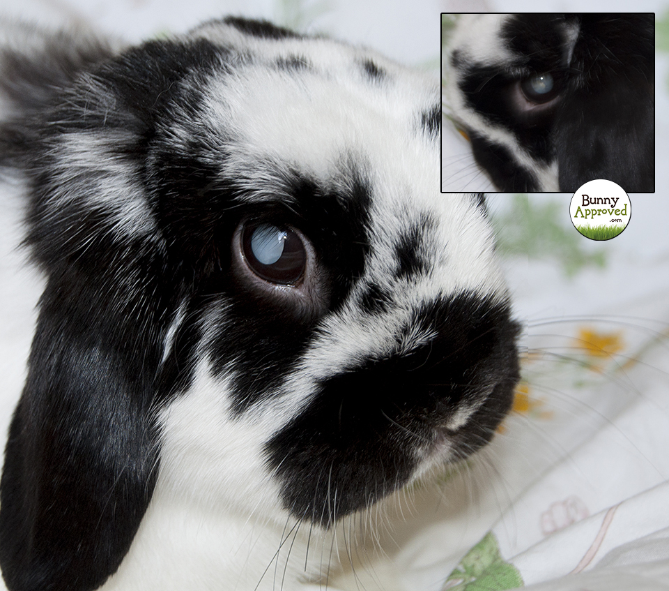 In December 2015 we noticed that Bunny’s left eye was looking a little hazy. At first we thought it was just a reflection from the window, but then it grew more cloudy and pronounced as the days went on. A visit to the vet confirmed that Bunny had developed a cataract. What is a cataract? Dr. Walters from NC State Veterinary Hospital explained it as follows in Bunny’s discharge papers:
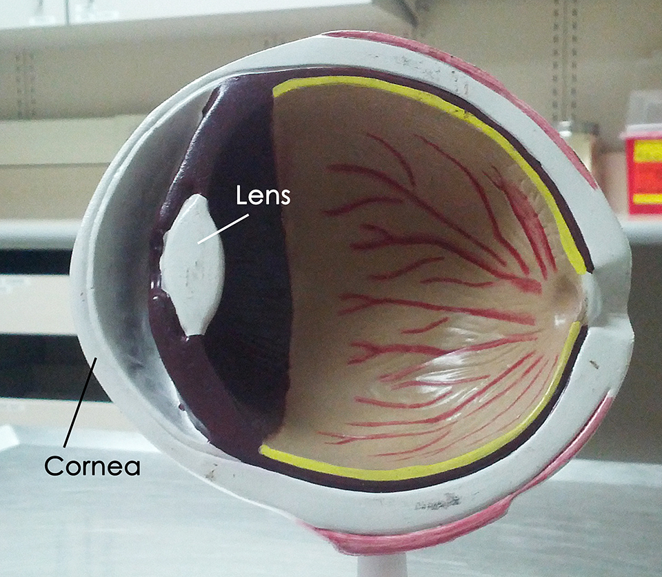
“Cataracts are an opacity of the lens, which is normally a clear, transparent structure that allows light to pass through the pupil to the retina in the back of the eye. The retina then transmits sensory visual information to the brain, which perceives this information as images. Under normal circumstances, the proteins in the lens are arranged in a highly regular fashion. Cataracts are caused by aggregation, or clumping, of the lens proteins. This clumping causes the lens to become opaque, preventing light from passing through and reaching the retina. This essentially renders the animal blind.”
Bunny’s eye quickly went from slightly hazy to completely cloudy in a short period of time. Dr. Walters explained that “cataracts are classified as incipient, immature, mature, or hypermature based on the percentage of the lens that is involved. Incipient cataracts are the earliest stage, and involve no more than 10-15% of the lens. Immature cataracts obscure between 15-99% of the lens. Patients with either of those two stages retain some degree of vision.”
Cataracts can develop at any time for no reason at all. They may be hereditary. Unfortunately, they can also be caused by E. Cuniculi, a parasite that many rabbits have been exposed to and that is often kept under control by the immune system only to take over as soon as the rabbit becomes sick, weak, or stressed. The test for EC is not particularly reliable, so even though Bunny tested negative for it, we still gave him Panacur for a few days to be sure.
Bunny’s behavior didn’t change much at all. He still had one good eye and losing sight in the other one did not prevent him from jumping, exploring, or finding his treats. Even though we knew about a possible surgery, we decided against it at that point because every surgery has its risks and Bunny’s quality of life seemed the same despite him being blind in one eye.
A few months passed before a second cataract began to develop in early May 2016. It only took about a week before Bunny was more or less blind in both eyes. He didn’t have much time to prepare and spent most of the day sitting in one spot, napping. He seemed angry, didn’t enjoy outdoor playtime anymore, and stopped exploring the house. He ran into things, got lost a couple of times, and heavily leaned on Bailey to show him the way. It took a good 3-4 weeks before he seemed to accept his blindness and became more confident around the house. We noticed that certain furniture and flooring helped him figure out where he was. The fleece blanket always had to stay on the floor, because it told him he was between the couch and his bench. The water bowl had to be placed in exactly the right spot, so it could lead him to the litter box. And whenever there was food, he would listen for Bailey’s chewing noises to find his share. Here’s a video we took a week before his surgery. He was extremely excited about the food, but simply couldn’t find it, despite my attempt to give him clues.
Finding a Vet
In May 2016 Bunny was only 6 years old. That’s way too young to be permanently blind if you have a choice! Even though Bunny was adapting to the new situation and could have led a nice life even while blind, we decided to go for the surgery. Unfortunately, there are not many Ophthalmologists who will perform surgery on rabbits. Our regular local vet contacted several animal hospitals in the area and none were willing to operate on Bunny. The closest one to us was nearly 3 hours away in Raleigh, NC. We didn’t know anything specific about the surgery and also didn’t know if Bunny would even qualify (for example, if the retinas are damaged, removing the cataracts will not restore vision), but we made an appointment for an exam on Tuesday, June 14th 2016. The surgery would be a day later and Bunny would have to stay until Thursday for observation. Ophthalmologists will need a referral from a regular vet, so even though you can find one online and contact them to see if they will treat rabbits, a regular vet will have to make the appointment for you after a general exam.
The Initial Exam
After the 3 hour car ride, we arrived at NC State Veterinary Hospital. The female human, Bunny, and Bailey. At first I had to answer questions about Bunny; how he lived, what he ate, and what kind of medical issues he had in the past. Then the hospital’s small animal vet, Dr. Walters, came in to take Bunny to the Ophthalmologist, Dr. Westermeyer. I was not present during the exam, but Dr. Walters explained it to me in the discharge papers as follows:
“Bunny was diagnosed with bilateral mature cataracts. […] Prior to surgery, Bunny had an ophthalmology consult to assess whether his eyes were otherwise normal and should be visual following cataract removal. A full ophthalmic exam was performed to evaluate for any signs of inflammation in the anterior chamber closest to the front of the eye. Because we could not see through the cataract to visually examine the back of the eye, we performed an electroretinogram in order to determine whether his retinas were functioning properly. Finally, an ocular ultrasound was performed to assess the integrity of the structures within Bunny’s eye. The results of these tests were normal.”
That meant that following a successful cataract surgery, Bunny should be able to see again.
The Risks
Any surgery has its risks. Rabbits don’t always do well under anesthesia and there is the possibility of death, either during the surgery or due to stasis after. However, an eye surgery is not as invasive as a spay, for example. Also, Bunny was healthy overall. He was at a good weight and had no other health issues that could have put him at a greater risk. If he had been weakened by E. Cuniculi, for example, we may have decided against the surgery.
Dr. Walters, the exotic vet, was present during the surgery to monitor Bunny and take action to keep him alive if necessary. She assured me that Dr. Westermeyer was very knowledgeable and had performed countless cataract surgeries on all kinds of animals, including rabbits. It made us feel good that someone was there who specializes in exotics to keep an eye on Bunny while an eye specialist worked on his cataracts.
Even though the surgery had its risks, we were informed that doing nothing was potentially risky as well:
“With time, the lens proteins can begin to leach out of the capsule surrounding the lens. At this stage, the cataract is referred to as hypermature. Having leaked out of the lens capsule and being exposed to the immune system, these lens proteins induce inflammation within the eye, known as uveitis, which is a painful condition. Other potential consequences of cataracts include glaucoma, a painful condition characterized by increased pressure within the eye, and detachment of the retina from the back of the eye, which results in permanent blindness that cannot be surgically corrected. For this reason, performing a phacoemulsification (surgery to remove the cataracts) is often recommended in the case of mature cataracts. This hopefully will restore Bunny’s vision without adverse effects.”
The Surgery
On the day of the surgery I dropped Bunny and Bailey off around 8am. The hospital allowed Bailey to stay to keep her sane (she doesn’t do well when they are separated) and support Bunny. The surgery took about 1-2 hours and Bunny was able to see immediately after waking up. In the doctor’s words:
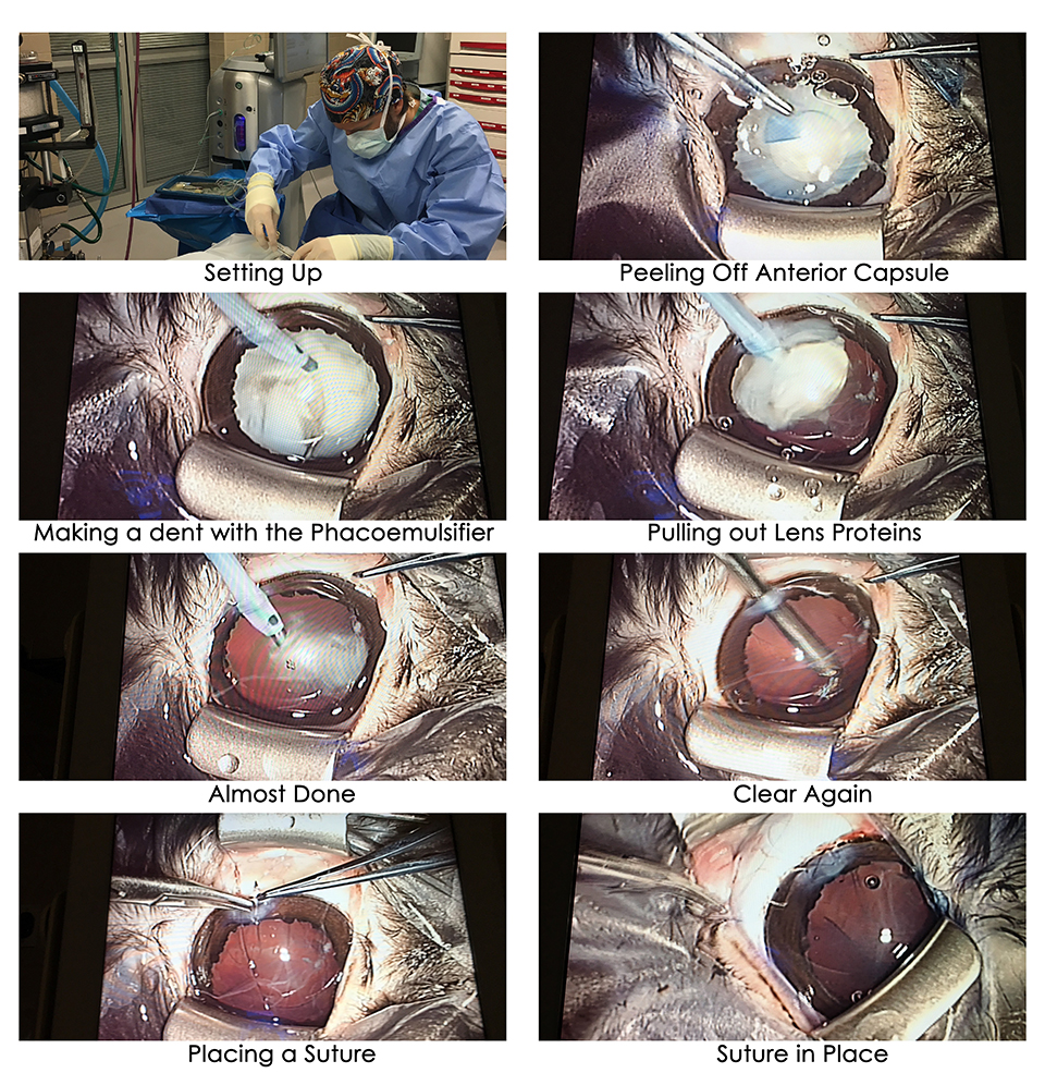
“Bunny’s cataracts were removed with a surgery called phacoemulsification. This employs a special tool that emits ultrasonic vibrations to liquefy the lens, and then extracts it with suction. In other species, the removed lens is typically replaced with an artificial lens. However, artificial lenses are not available in suitable sizes for rabbits. The lens normally adjusts in shape to allow the eye to focus on objects at different distances, a process called accommodation. Without that lens, Bunny will not be able to focus; as a result, Bunny’s vision will be slightly blurry without his lens. However, rabbits do not have a very good capacity for accommodation to begin with, so this change may not be very noticeable or significant for Bunny, and he should be able to adapt well over time. Now that the opaque lens material has been removed from his eyes and his retina is functioning normally, Bunny should be able to see. You may notice small scars with tiny sutures where the surgical incisions were made on the upper aspect of Bunny’s eyes at the junction between the white part of his eye (the sclera) and the colored portion of the eye (the iris). The sutures will dissolve and will not need to be specifically removed. Bunny may have a few red blood vessels on his sclera, which is part of the healing process of the eye and should resolve over the course of a few days. The pink mucous membranes on the underside of his eyelids, known as the conjunctiva, may also appear slightly more red than usual for a few days.”
 Dr. Walters also told me that due to the incision, the fluid between the cornea and the lens was drained, so they added new fluid towards the end of the surgery. Over time, Bunny will regenerate his own, but until then it could form visible bubbles, which is normal and no cause for concern.
After the Surgery
Once Bunny had woken up from the surgery, he was allowed to stay with Bailey in a cage at the hospital. He was sleepy and still had a catheter attached to his leg with some Vetrap bandages to make it easier in case he needed pain medication. I went to visit a few hours later and he was still very sleepy, but his eyes looked normal again and he could see me! Both bunnies were discharged on Thursday and we went back home. Bunny didn’t eat much that day and he seemed to be in a little bit of pain from the catheter, so I gave him some Metacam. He perked up a little and was back to normal the next day. He is now as active as he was before the cataracts developed and hasn’t had any problems seeing his environment.
We were asked to come back for a check-up one week after the surgery and three weeks after the surgery. There is a vet in Greensboro that can do the check-up, so we don’t have to drive all the way to Raleigh this time. The one right here in town (not our regular vet) was too busy to see us this month. Looking back, it would have been good to schedule a visit with them earlier. Anyway, during those three weeks we have to give Bunny the following eye-drops:
- Diclofenac: Administer 1 drop in each eye every 6 hours (four times a day) until directed otherwise.
- Ciprofloxacin: Administer 1 drop in each eye every 6 hours (four times a day) until directed otherwise.
- Tropicamide: Administer 1 drop in each eye every 12 hours (twice a day) until directed otherwise.
That’s 10 eye drops a day in each eye! Not the most pleasant thing to have to do when your rabbit tries to find new ways to avoid them every single day, but easier than syringe-feeding. It’s also quite a time commitment. In Bunny’s discharge papers Dr. Walters added that his “ciprofloxacin eye drops contain an antibiotic to help prevent an infection from occurring, and the tropicamide eye medication dilates the pupil to aid intraocular healing and help prevent adhesions from forming between the iris and the cornea (anterior synechiae) or between the iris and the lens (posterior synechiae). When administering Bunny’s eye drops, please wait 5 minutes after giving one medication before giving the next to allow the first drop to be absorbed.”
Costs
We were quoted between $1800 and $2200 for the surgery including all the exams and the hospital stay. We ended up paying $1656 for everything that happened during our 3 days in Raleigh. Hotel and gas not included. We have not gone back yet for the follow-up exams, but will update the post later.
UPDATE:
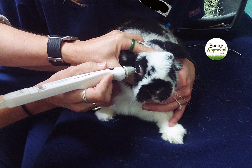 Bunny went to an ophthalmologist in Greensboro who graciously agreed to check his eyes one week after the surgery. That way we only had about 90min. to drive. The check-up took about 10 minutes and his incision looked perfect. He had zero inflammation and everything was healing well. The doctor also performed a tonometry, which means she measured the eye pressure to make sure Bunny hadn’t developed a glaucoma. Luckily, he hadn’t! We were able to reduce the amount of eye drops to 2 different medications 3 times a day.
At this point we needed a refill on the Diclofenac. The Greensboro vet didn’t have any in stock, so she gave us a prescription to be filled by any pharmacy. CVS charged us almost $75 for the exact same bottle we had at home. When I checked our hospital bill later, it said we only paid about $17 for the first bottle. Lesson learned! All future refills will be ordered from the hospital that did Bunny’s eye surgery!
Another two weeks later we went for our second check-up. Bunny’s eyes were healing exactly the way they should! We were now down to 1 eye drop a day (our overpriced Diclofenac). Several check-ups followed with more and more time in between. It’s now 8 months post surgery and Bunny is doing well. He can see without any noticeable issues and has not developed a glaucoma or an infection.
UPDATE 2: Around 9 months after the surgery Bunny’s eye pressure levels increased slightly. They are not above normal, but on the higher end of normal. In order to prevent a glaucoma, Bunny was prescribed Dorzolamide 2% eye drops that he needs twice a day. The eye doctor said he may have to be on these long-term. They cost $31.92 when we purchased them from the vet and $85 at a local Rite Aid. Each bottle lasts about 6-7 weeks.
UPDATE 3: About 18 months have passed since the surgery and Bunny’s eye pressure has gone back to normal. He still gets Dorzolamide 2% eye drops, but only once a day as a precaution. We have finally found an animal eye clinic nearby that accepts rabbits, so we don’t have to drive all the way to Greensboro anymore. His check-ups are also down to twice a year now. Bunny is still happy and healthy overall! He can see well and the fact that he can’t focus anymore is only noticeable occasionally when he misjudges distances and jumps too far or bumps his head on things. This really is a rare thing, though!
Dr. Walters knew that I wanted to write about our experience and provided some awesome pictures and even a video of the procedure. They are not terribly gruesome (there’s no blood and you can’t really tell that it’s Bunny on the operating table), so if you want to see them, keep scrolling. If not, now is a good time to stop.
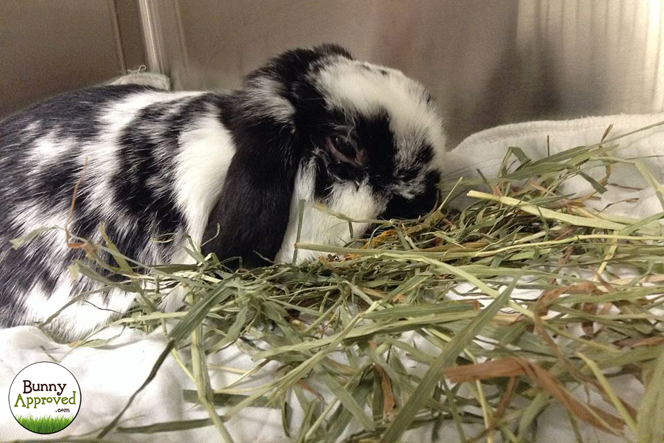
Bunny shortly after the surgery. He was still very sleepy.
Tags: approved, Blind, Bunny, Cataract, eye, eyes, mature, rabbit, See, surgery
Categories: All, Grooming & Health